Breast Pain or Lumps: What Tests Do You Need?
Last updated: 1 Aug 2026
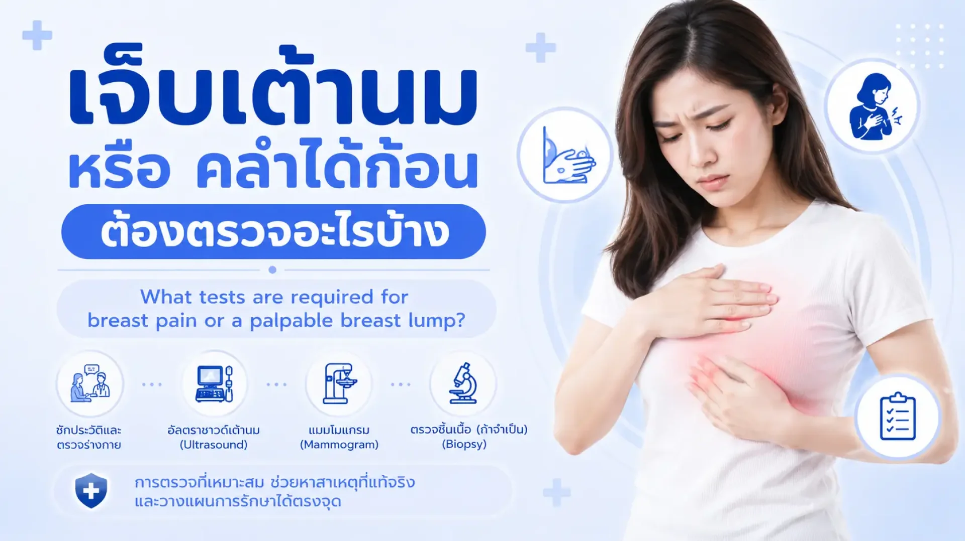
Breast Pain or Lumps: What Screenings and Tests Do You Need?
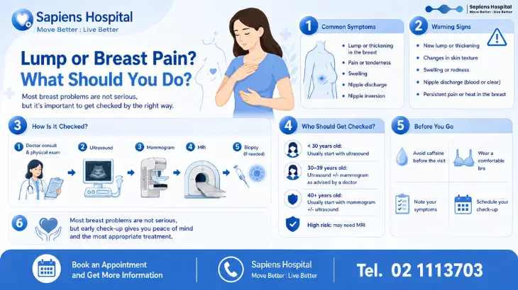
Experiencing breast pain or discovering a lump can cause immediate anxiety for many women, often prompting the thought, “Could this be breast cancer?” This concern is completely understandable, as breast cancer is common among women, and early detection significantly improves treatment outcomes. However, medically speaking, breast pain or a breast lump does not always indicate cancer. In many cases, these symptoms are caused by hormonal changes, breast cysts, benign tumors, inflammation, or natural age-related changes in breast tissue.
Nevertheless, knowing that "most cases are not cancer" does not mean you should skip getting checked. Certain types of lumps require monitoring, some may necessitate a biopsy, and specific symptoms are clear warning signs that warrant an immediate visit to a doctor.
This article will help you understand which types of breast pain are concerning, what tests are needed if you find a lump, how age affects the choice of screening, and when you should seek medical attention instead of waiting to see if symptoms improve.
What Causes Breast Pain?
Breast pain or tenderness is very common, especially in menstruating women. Many times, these symptoms are directly related to hormonal fluctuations during the menstrual cycle—such as experiencing pain or engorgement before a period, which resolves once menstruation begins.
Common causes include:
Which Types of Breast Pain Are Generally Less Concerning?
Breast pain associated with hormonal changes or benign conditions typically presents with the following characteristics:
When to See a Doctor
You should seek medical attention if you experience any of the following symptoms:
What Could a Breast Lump Be?
Breast lumps can stem from a variety of causes. Some are benign, some require ongoing observation, and others require a biopsy.
Evaluating breast pain or lumps involves more than just a physical examination. Doctors use a combination of assessments, factoring in age, symptoms, lump location, personal risk factors, and physical exam findings. The standard approach is known as the "Triple Assessment," which includes:
1. Medical History and Clinical Breast Exam
The doctor will ask detailed questions, such as:
2. Breast Ultrasound
A breast ultrasound uses sound waves (no radiation) and is ideal for younger women, those with dense breast tissue, or evaluating a lump in a specific location. It can accurately distinguish between fluid-filled cysts and solid masses, and identify features that may require a biopsy.
3. Mammogram
A mammogram is an X-ray of the breast. It is used both for screening (in women with no symptoms) and for diagnostic purposes (in individuals experiencing symptoms like a palpable lump, localized pain, nipple abnormalities, or skin changes). Mammograms can detect abnormalities that cannot be felt, such as microcalcifications or structural distortions.
4. Breast MRI
A breast MRI is not a first-line test for every patient but is highly beneficial in specific situations, such as:
5. Core Needle Biopsy
If imaging reveals suspicious characteristics or the nature of the lump remains unclear, a doctor will likely recommend a biopsy for pathological examination. This provides a definitive answer regarding whether the tissue is benign, requires monitoring, or is cancerous.
Many people worry that a biopsy will cause cancer to spread. However, medically speaking, a biopsy performed with proper clinical techniques is the standard for diagnosis and is essential for effective treatment planning.
How Age Affects Your Screening Plan
Breast screening approaches vary by age due to differences in breast tissue density and disease risk:
Red Flag Symptoms: When to Seek Immediate Care
Do not wait to see a doctor if you experience any of the following:
Breastfeeding Concerns: What to Watch Out For
Nursing mothers frequently experience breast engorgement, clogged milk ducts, or mastitis. Symptoms generally include a breast that is red, hot, painful, accompanied by a fever, or feeling flu-like. If not managed correctly, this can develop into a breast abscess. See a doctor if:
Do Men Need to Be Checked for Breast Lumps?
While breast cancer in men is rare compared to women, men can still develop breast lumps. Common causes include gynecomastia (enlarged breast tissue due to hormones, medications, liver disease, etc.). Male breast cancer, though uncommon, does occur. Men should see a doctor if they notice:
Screening vs. Diagnostic Testing: What’s the Difference?
Self-Care for Mild Breast Pain
If your breast pain is cyclical, affects both sides, and you have no lumps or red-flag symptoms, you can try these initial self-care steps:
Frequently Asked Questions (FAQs)
Does breast pain mean I have cancer?
Not necessarily. Most breast pain is linked to hormones, cysts, inflammation, or benign causes. However, if the pain is strictly localized, continuous, one-sided, or accompanied by a lump, nipple changes, or skin abnormalities, you should see a doctor.
What tests are needed if I find a breast lump?
Evaluation typically begins with a clinical breast exam by a doctor, followed by imaging such as an ultrasound or mammogram (depending on age and the lump's characteristics). If the results are suspicious, a biopsy may be required for confirmation.
Do I need a mammogram if I am young?
For women under 30 with a lump, an ultrasound is usually the first step. If the ultrasound is suspicious or if the patient is at high genetic risk, a doctor may recommend a mammogram or additional imaging.
Is it true that painful lumps are usually not cancer?
Painful lumps are frequently caused by cysts, inflammation, or benign conditions. However, this does not rule out cancer entirely. Any new, growing, or unusual lump should be clinically evaluated.
Is bleeding from the nipple dangerous?
You should see a doctor immediately, especially if the bleeding is from one side, occurs spontaneously without squeezing, or is continuous. It could indicate abnormal milk ducts, intraductal papillomas (benign polyps), or conditions requiring further evaluation.
What is the difference between an ultrasound and a mammogram?
An ultrasound uses sound waves (no radiation) and is excellent for evaluating specific lumps and fluid-filled cysts, especially in younger women or those with dense breasts. A mammogram uses X-rays to detect structural abnormalities and microcalcifications, and is the standard for breast cancer screening in the appropriate age groups.
What should I do if my mammogram is normal but I can still feel a lump?
You should follow up with your doctor. In some cases, a supplementary ultrasound or a biopsy may be necessary if a distinct lump is palpable but imaging results do not provide a clear answer.
Breast pain and lumps are common symptoms and are frequently not cancer. However, relying solely on self-examination or how the lump "feels" is not enough. If your pain fluctuates with your cycle, affects both breasts, and has no other abnormalities, it may be a benign condition. But if you find a new lump, experience continuous localized pain, nipple bleeding, skin dimpling, shape changes, or an armpit lump, you should consult a physician to be thoroughly evaluated.
The most important takeaway: Don't panic, but don't wait too long either. Prompt medical evaluation provides clear answers, allows for accurate treatment planning, and ensures that if a serious condition is present, it can be managed effectively in its earliest stages.
For Inquiries and Appointments Sapiens Hospital | Move Better : Live Better Tel. 02-111-3703
References
Klein KA, Kocher M, Lourenco AP, et al. ACR Appropriateness Criteria® Palpable Breast Masses. Journal of the American College of Radiology. 2023.
Holbrook AI, Moy L, Akin EA, et al. ACR Appropriateness Criteria® Breast Pain. Journal of the American College of Radiology. 2018.
U.S. Preventive Services Task Force. Screening for Breast Cancer: US Preventive Services Task Force Recommendation Statement. JAMA. 2024;331(22):1918–1930.
National Institute for Health and Care Excellence. Suspected cancer: recognition and referral. NICE Guideline NG12. Updated guidance.
National Comprehensive Cancer Network. NCCN Clinical Practice Guidelines in Oncology: Breast Cancer Screening and Diagnosis. Version updates.
Britton P. Breast imaging. BMJ. 2008;337:a1313.
Karim MO, Khan KA, Khan AJ, et al. Triple Assessment of Breast Lump: Should We Perform Core Biopsy for Every Patient? Cureus. 2020.
American College of Radiology. ACR BI-RADS® Atlas: Breast Imaging Reporting and Data System.
Experiencing breast pain or discovering a lump can cause immediate anxiety for many women, often prompting the thought, “Could this be breast cancer?” This concern is completely understandable, as breast cancer is common among women, and early detection significantly improves treatment outcomes. However, medically speaking, breast pain or a breast lump does not always indicate cancer. In many cases, these symptoms are caused by hormonal changes, breast cysts, benign tumors, inflammation, or natural age-related changes in breast tissue.
Nevertheless, knowing that "most cases are not cancer" does not mean you should skip getting checked. Certain types of lumps require monitoring, some may necessitate a biopsy, and specific symptoms are clear warning signs that warrant an immediate visit to a doctor.
This article will help you understand which types of breast pain are concerning, what tests are needed if you find a lump, how age affects the choice of screening, and when you should seek medical attention instead of waiting to see if symptoms improve.
What Causes Breast Pain?
Breast pain or tenderness is very common, especially in menstruating women. Many times, these symptoms are directly related to hormonal fluctuations during the menstrual cycle—such as experiencing pain or engorgement before a period, which resolves once menstruation begins.
Common causes include:
- Hormonal changes throughout the menstrual cycle
- Breast cysts
- Dense breast tissue or fibrocystic breast changes
- Breast inflammation (mastitis)
- Breastfeeding or clogged milk ducts
- Chest trauma or chest wall muscle inflammation
- Certain medications or hormone therapies
- Stress, lack of sleep, high caffeine intake, or lifestyle triggers in some individuals
Which Types of Breast Pain Are Generally Less Concerning?
Breast pain associated with hormonal changes or benign conditions typically presents with the following characteristics:
- Pain spreading across both breasts or a wide area
- Tenderness or engorgement prior to menstruation
- Symptoms improve after menstruation begins
- No distinct, palpable lump
- No bleeding or abnormal discharge from the nipples
- No skin dimpling, redness, thickening, or changes in breast shape
- Symptoms come and go with the menstrual cycle
When to See a Doctor
You should seek medical attention if you experience any of the following symptoms:
- Continuous breast pain localized to one specific spot
- Pain in only one breast that is unrelated to your menstrual cycle
- A newly discovered, palpable lump
- Nipple inversion, retraction, or changes in shape
- Clear fluid or blood discharging from the nipple (especially from one side)
- Skin on the breast that is dimpled, puckered, red, thickened, or resembles an orange peel (peau d'orange)
- Breast swelling, redness, heat, severe pain, or fever
- A palpable lump in the armpit
- A family history of breast or ovarian cancer
- Being over the age of 40 and experiencing new, unfamiliar symptoms
What Could a Breast Lump Be?
Breast lumps can stem from a variety of causes. Some are benign, some require ongoing observation, and others require a biopsy.
- Breast Cysts: Fluid-filled sacs that are very common, particularly in women aged 30–50. The lumps are usually round and smooth, and may become more tender before a period. An ultrasound can easily distinguish a fluid-filled cyst from a solid mass.
- Fibroadenoma: A common, benign (non-cancerous) tumor frequently found in younger women. These lumps are typically round, have smooth borders, are easily movable, and grow slowly. In some cases, a doctor may recommend monitoring or a biopsy to confirm the diagnosis.
- Breast Infection or Mastitis: Often found in lactating women or those with clogged milk ducts. Symptoms usually include redness, heat, pain, and fever, and can sometimes lead to a breast abscess.
- Fat Necrosis: Some individuals develop fat necrosis after breast trauma or surgery, resulting in a hard lump that needs to be medically distinguished from other conditions.
- Breast Cancer: A cancerous lump often feels hard, has irregular borders, is fixed to the surrounding tissue, and is usually painless. It may also be accompanied by changes to the skin, nipple, or armpit. Because symptoms can sometimes be subtle, any new breast lump should be evaluated by a doctor.
Evaluating breast pain or lumps involves more than just a physical examination. Doctors use a combination of assessments, factoring in age, symptoms, lump location, personal risk factors, and physical exam findings. The standard approach is known as the "Triple Assessment," which includes:
- Clinical Assessment: Medical history and clinical breast examination by a doctor.
- Imaging: Breast imaging (e.g., Ultrasound or Mammogram).
- Tissue Biopsy: Core needle biopsy or fine-needle aspiration (if imaging results are suspicious or inconclusive).
1. Medical History and Clinical Breast Exam
The doctor will ask detailed questions, such as:
- When did the symptoms start?
- Is the pain on one side or both?
- Is the pain related to your menstrual cycle?
- How long have you felt the lump?
- Has the lump grown or changed?
- Is there any discharge or bleeding from the nipple?
- Do you have a personal history of breast lumps or surgeries?
- Is there a family history of breast or ovarian cancer?
- Are you taking oral contraceptives, hormone replacement therapy, or other hormones?
- Are you currently pregnant, breastfeeding, or have you recently stopped breastfeeding?
2. Breast Ultrasound
A breast ultrasound uses sound waves (no radiation) and is ideal for younger women, those with dense breast tissue, or evaluating a lump in a specific location. It can accurately distinguish between fluid-filled cysts and solid masses, and identify features that may require a biopsy.
- For women under 30: An ultrasound is generally the first imaging test used when a lump is found.
- Used to evaluate specific, palpable lumps.
- Used to assess cysts, abscesses, or solid tumors.
- Used alongside a mammogram in older women.
- Used to guide the needle during a biopsy if necessary.
3. Mammogram
A mammogram is an X-ray of the breast. It is used both for screening (in women with no symptoms) and for diagnostic purposes (in individuals experiencing symptoms like a palpable lump, localized pain, nipple abnormalities, or skin changes). Mammograms can detect abnormalities that cannot be felt, such as microcalcifications or structural distortions.
- Women aged 40 and older should discuss regular screening mammograms with their doctor.
- If a lump is found in a woman over 40, a diagnostic mammogram is typically performed in conjunction with an ultrasound.
- For women aged 30–39, doctors may recommend an ultrasound, a mammogram, or both, depending on symptoms and risk factors.
- A diagnostic mammogram differs from a screening mammogram because it focuses specifically on evaluating an identified symptom or localized abnormality.
4. Breast MRI
A breast MRI is not a first-line test for every patient but is highly beneficial in specific situations, such as:
- Individuals at high risk for breast cancer.
- Individuals with a genetic predisposition (e.g., BRCA1/BRCA2 mutations).
- Those with extremely dense breasts and specific clinical indications.
- Evaluating the extent of the disease in patients already diagnosed with breast cancer.
- Cases where other imaging results are inconclusive and further clarity is required.
5. Core Needle Biopsy
If imaging reveals suspicious characteristics or the nature of the lump remains unclear, a doctor will likely recommend a biopsy for pathological examination. This provides a definitive answer regarding whether the tissue is benign, requires monitoring, or is cancerous.
Many people worry that a biopsy will cause cancer to spread. However, medically speaking, a biopsy performed with proper clinical techniques is the standard for diagnosis and is essential for effective treatment planning.
How Age Affects Your Screening Plan
Breast screening approaches vary by age due to differences in breast tissue density and disease risk:
- Under 30 Years Old: If a lump is found, an ultrasound is usually the first step. Younger breast tissue is typically dense, and benign conditions like fibroadenomas or cysts are much more common. If ultrasound results are suspicious, further tests or a biopsy may follow.
- Ages 30–39: Depending on the symptoms, lump location, and personal risk, doctors may utilize an ultrasound, a diagnostic mammogram, or both to gather the most accurate information.
- Age 40 and Older: For a palpable lump, a diagnostic mammogram combined with an ultrasound is the standard approach. The mammogram evaluates the entire breast and can spot abnormalities that an ultrasound alone might miss.
Red Flag Symptoms: When to Seek Immediate Care
Do not wait to see a doctor if you experience any of the following:
- A new lump in the breast or armpit.
- A lump that is hard, has irregular borders, or feels fixed/immovable.
- A lump that is continuously growing.
- Breast skin that is dimpled, puckered, thickened, red, or resembles an orange peel.
- A nipple that turns inward (inverted), retracts, or changes direction.
- Clear fluid or blood discharging from the nipple, especially on one side.
- A noticeable change in the size or shape of the breast.
- A sore or rash on the nipple that does not heal.
- A breast that is red, hot to the touch, highly painful, and accompanied by a fever.
- Continuous, localized breast pain unrelated to the menstrual cycle.
- You have a strong family history of breast or ovarian cancer.
Breastfeeding Concerns: What to Watch Out For
Nursing mothers frequently experience breast engorgement, clogged milk ducts, or mastitis. Symptoms generally include a breast that is red, hot, painful, accompanied by a fever, or feeling flu-like. If not managed correctly, this can develop into a breast abscess. See a doctor if:
- You have a high fever.
- The breast is extremely red and painful.
- You feel a painful lump that does not go down after feeding or pumping.
- There is pus or an open wound.
- Symptoms do not improve within 24–48 hours.
- The pain is so severe that you cannot breastfeed.
Do Men Need to Be Checked for Breast Lumps?
While breast cancer in men is rare compared to women, men can still develop breast lumps. Common causes include gynecomastia (enlarged breast tissue due to hormones, medications, liver disease, etc.). Male breast cancer, though uncommon, does occur. Men should see a doctor if they notice:
- A hard lump underneath the nipple or on the side of the breast.
- A lump that is growing.
- Nipple inversion or bleeding.
- Skin abnormalities.
- A lump in the armpit.
- Noticeable swelling or pain on one side.
Screening vs. Diagnostic Testing: What’s the Difference?
- Screening is for individuals with no symptoms, aimed at catching diseases in their earliest stages (e.g., an annual screening mammogram).
- Diagnostic Workup is for individuals who have symptoms (e.g., a palpable lump, bloody nipple discharge, skin dimpling, or continuous localized pain). This type of testing is specifically tailored to diagnose the cause of the symptom, rather than being a general screening.
Self-Care for Mild Breast Pain
If your breast pain is cyclical, affects both sides, and you have no lumps or red-flag symptoms, you can try these initial self-care steps:
- Wear a well-fitting, supportive bra and avoid excessive pressure on the breasts.
- Track the pain to see if it correlates with your menstrual cycle.
- Reduce caffeine intake if you notice it triggers your symptoms.
- Ensure you get adequate sleep and rest.
- Take over-the-counter pain relievers as advised by a doctor or pharmacist.
- Keep a record of exactly where and when the pain occurs.
Frequently Asked Questions (FAQs)
Does breast pain mean I have cancer?
Not necessarily. Most breast pain is linked to hormones, cysts, inflammation, or benign causes. However, if the pain is strictly localized, continuous, one-sided, or accompanied by a lump, nipple changes, or skin abnormalities, you should see a doctor.
What tests are needed if I find a breast lump?
Evaluation typically begins with a clinical breast exam by a doctor, followed by imaging such as an ultrasound or mammogram (depending on age and the lump's characteristics). If the results are suspicious, a biopsy may be required for confirmation.
Do I need a mammogram if I am young?
For women under 30 with a lump, an ultrasound is usually the first step. If the ultrasound is suspicious or if the patient is at high genetic risk, a doctor may recommend a mammogram or additional imaging.
Is it true that painful lumps are usually not cancer?
Painful lumps are frequently caused by cysts, inflammation, or benign conditions. However, this does not rule out cancer entirely. Any new, growing, or unusual lump should be clinically evaluated.
Is bleeding from the nipple dangerous?
You should see a doctor immediately, especially if the bleeding is from one side, occurs spontaneously without squeezing, or is continuous. It could indicate abnormal milk ducts, intraductal papillomas (benign polyps), or conditions requiring further evaluation.
What is the difference between an ultrasound and a mammogram?
An ultrasound uses sound waves (no radiation) and is excellent for evaluating specific lumps and fluid-filled cysts, especially in younger women or those with dense breasts. A mammogram uses X-rays to detect structural abnormalities and microcalcifications, and is the standard for breast cancer screening in the appropriate age groups.
What should I do if my mammogram is normal but I can still feel a lump?
You should follow up with your doctor. In some cases, a supplementary ultrasound or a biopsy may be necessary if a distinct lump is palpable but imaging results do not provide a clear answer.
Breast pain and lumps are common symptoms and are frequently not cancer. However, relying solely on self-examination or how the lump "feels" is not enough. If your pain fluctuates with your cycle, affects both breasts, and has no other abnormalities, it may be a benign condition. But if you find a new lump, experience continuous localized pain, nipple bleeding, skin dimpling, shape changes, or an armpit lump, you should consult a physician to be thoroughly evaluated.
The most important takeaway: Don't panic, but don't wait too long either. Prompt medical evaluation provides clear answers, allows for accurate treatment planning, and ensures that if a serious condition is present, it can be managed effectively in its earliest stages.
For Inquiries and Appointments Sapiens Hospital | Move Better : Live Better Tel. 02-111-3703
References
Klein KA, Kocher M, Lourenco AP, et al. ACR Appropriateness Criteria® Palpable Breast Masses. Journal of the American College of Radiology. 2023.
Holbrook AI, Moy L, Akin EA, et al. ACR Appropriateness Criteria® Breast Pain. Journal of the American College of Radiology. 2018.
U.S. Preventive Services Task Force. Screening for Breast Cancer: US Preventive Services Task Force Recommendation Statement. JAMA. 2024;331(22):1918–1930.
National Institute for Health and Care Excellence. Suspected cancer: recognition and referral. NICE Guideline NG12. Updated guidance.
National Comprehensive Cancer Network. NCCN Clinical Practice Guidelines in Oncology: Breast Cancer Screening and Diagnosis. Version updates.
Britton P. Breast imaging. BMJ. 2008;337:a1313.
Karim MO, Khan KA, Khan AJ, et al. Triple Assessment of Breast Lump: Should We Perform Core Biopsy for Every Patient? Cureus. 2020.
American College of Radiology. ACR BI-RADS® Atlas: Breast Imaging Reporting and Data System.
Related Content

หากล้มหัวฟาดพื้นและปวดคอ ให้สังเกตอาการ เช่น ความเจ็บปวดที่รุนแรงหรือความชาบริเวณคอ ถ้ามีอาการเหล่านี้ ควรรีบไปพบแพทย์เพื่อรับการรักษาที่เหมาะสม
28 May 2025
The word 'tumor' often causes alarm, as many fear it means cancer. However, most uterine fibroids are benign, non-cancerous, and do not always require surgery.
9 Jul 2026
